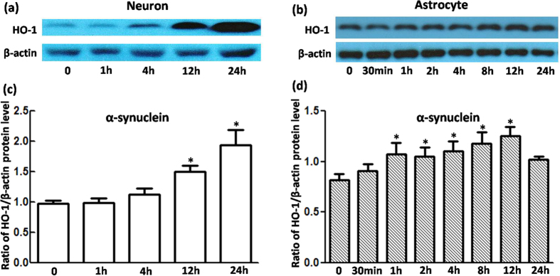
Figure 2. HO-1 protein levels were upregulated in VM neurons and astrocytes treated with α-synuclein.
(a,b) Western blots were used to detect HO-1 protein levels in cells with 5 μg/mL α-synuclein treatment. HO-1 expression in neurons increased at 12 h and 24 h. For astrocytes, HO-1 upregulation was detected at 1 h, 2 h, 4 h, 8 h, 12 h, and 24 h. β-Actin was used as a loading control. (c,d) Statistical analysis. The data are presented as the ratio of HO-1 to β-actin. Each bar represents the mean ± S.E.M. of more than 6 independent experiments. *P < 0.05, compared with control.

