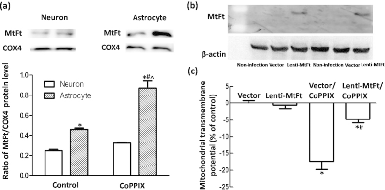
Figure 9. MtFt overexpression rescued ΔΨm collapse in neurons with CoPPIX administration.
(a) Western blots were used to detect MtFt protein levels in neurons and astrocytes with CoPPIX administration for 24 h. MtFt levels were higher and more robustly upregulated in astrocytes than in neurons. The data are presented as the ratio of MtFt to COX4. Each bar represents the mean ± S.E.M. of more than 6 independent experiments. *P < 0.05, compared with neurons without CoPPIX treatment; #P < 0.05, compared with astrocytes without CoPPIX treatment; ^P < 0.05, compared with neurons with CoPPIX treatment. (b) Western blots were applied to detect MtFt protein levels in neurons with pLenti-Ubc-MtFt or pLenti-Ubc-MCS transduction. (c) Flow cytometry was applied to detect ΔΨm in different groups. Lentiviral transduction (pLenti-Ubc-MtFt or pLenti-Ubc-MCS) had no effect on ΔΨm. CoPPIX (25 μmol/L) induced ΔΨm reduction in the lentiviral vector group. However, a significant restoration was observed in the MtFt over-expression group. The data are presented as the mean ± S.E.M. of more than 4 independent experiments. *P < 0.05, compared with lentivector group; #P < 0.05, compared with lentivector/CoPPIX group.

