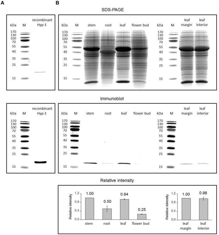
FIGURE 4.
Immunoblotting detection of Hyp-1 protein in H. perforatum tissues. Coomassie Brilliant Blue stained SDS-PAGE gels and the corresponding immunoblots showing detection of Hyp-1 protein in samples of recombinant Hyp-1 protein (A) and H. perforatum tissues (B). Lane M, protein molecular mass marker, with size (kDa) indicated on the left. Relative intensity values for protein levels in immunoblots represent means ± SE of three biological replicates.

