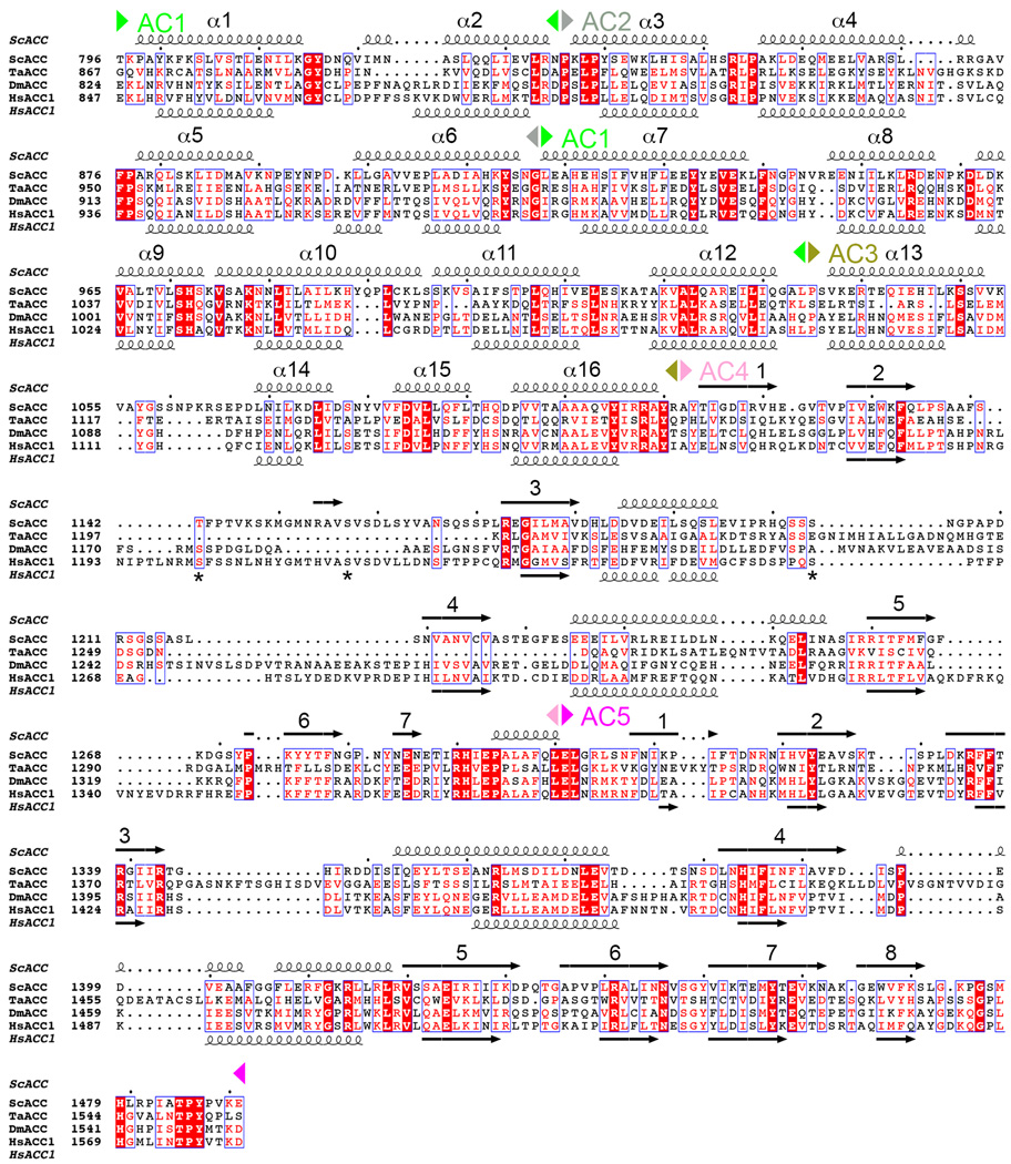
Extended Data Fig. 2.
Sequence alignment of the central region of eukaryotic, single-chain ACCs. The AC1–5 domains are indicated. Predicted secondary structure elements in HsACC1 are also shown, and they generally match those in the structure of ScACC. The helices in AC1–3 are numbered consecutively. Three sites of phosphorylation in HsACC1 are indicated with stars.

