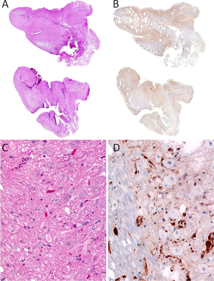
Fig. 4.
Dysplastic cerebellar gangliocytoma; patient with Lhermitte–Duclos. a, c H&E sections, b, c PTEN IHC immunostain. a Shows folial expansion of cerebellum, where the darkly staining granular cell layer is lost. c Shows the folial expansion corresponds to reduced PTEN expression by IHC. b Shows the granular cell layer being replaced by ganglion cells of varying sizes. d Shows many of the ganglion cells have lost PTEN expression. Note the retention of PTEN expression in background vasculature

