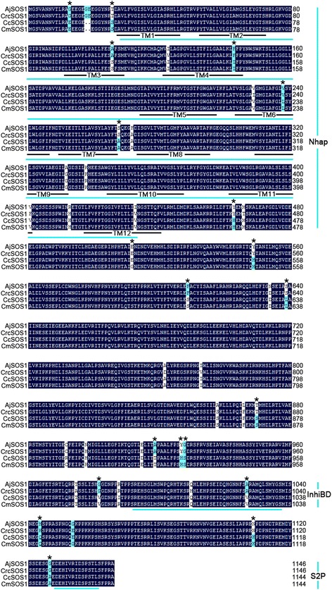
Fig. 3.

Multiple amino acid sequence alignment between four SOS1s. The 12 putative transmembrane domains are underlined and numbered 1 through 12. Residues conserved in at least two proteins are highlighted in white and blue. The black asterisks indicate conserved residues which were replaced in the site-directed mutagenesis experiment (see Additional file 2: Figure S4). Nhap, an Na+/H+ exchanger domain spanning the transmembrane region; InhiBD, an auto-inhibitory domain; S2P, SOS2 phosphorylation motif
