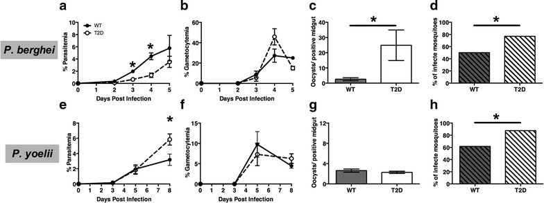
Fig. 1.

Parasitaemias, gametocytaemias and parasite transmission from wild-type (WT) and type 2 diabetic (T2D) C57BL/6 mice infected with malaria. a, e Parasitaemias and b, f gametocytaemias (mean ± SEM) were determined by visual counts from Giemsa-stained slides prepared from tail bleeds. n = 4–6, *p ≤ 0.05). c, d, g, h Prior to peak parasitaemia (day 5 for Plasmodium berghei and day 8 for Plasmodium yoelii yoelii 17XNL), mice were anesthetized and 50 mosquitoes/mouse were allowed to feed for 15 min. Mosquitoes were maintained on 10 % sucrose for 10 days at which point midguts were dissected and oocysts counted. n = 30–70 mosquitoes, *p ≤ 0.05
