Fig. 3.

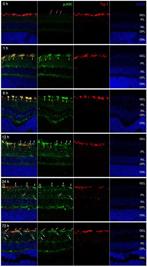
Phosphorylated JNK was detected in retina after I/R injury. Frozen-sectioned (10 μm) retina samples from 0, 1, 6, 12, 24 and 72 h after I/R injury were used for immunohistochemistry. Phosphorylated JNK was detected (green fluorescence). Tuj-1 immunofluorescence (red) was used as RGC marker. DAPI staining (blue) represents cell nuclei for counter staining. The magenta arrows represent basal JNK phosphorylation in 0 h retina. All yellow arrows represent phosphorylated JNK in RGC and white arrows represent non-RGC in GCL
