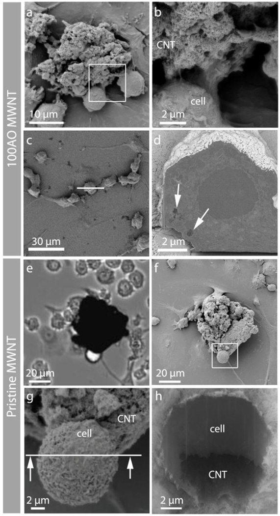
Figure 3.
Correlative microscopy showing the interaction between 100AO and pristine MWNT aggregates and N9 microglia. (a,b) Low and high magnification SEM micrographs showing N9 microglia interacting with an 100AO MWNT aggregate. (c) Top view of another N9 microglia, which was sectioned by FIB milling at the white line. (d) Back scattered electron (BSE) image of the cross-sectioned surface reveals the presence of sub-micron aggregates of 100AO MWNT inside the microglia (arrows). (e) Light micrograph of pristine MWNT aggregate and (f) corresponding SEM images of the same area after critical point drying at low (g) and higher (h) magnification. The partial wrapping of the microglia around part of the aggregate demonstrates the clearance challenge that large pristine MWNT aggregates presents to the cells.

