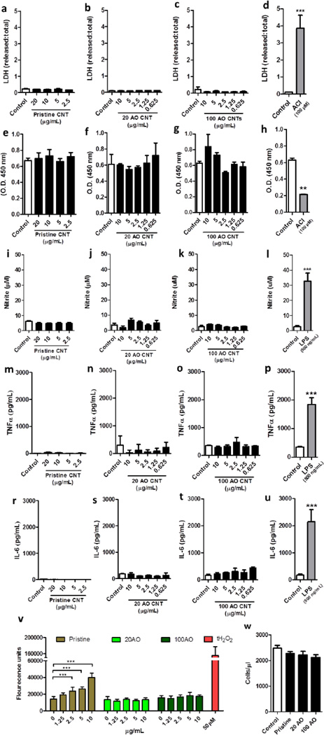
Figure 5.
Microglial viability and inflammatory response after exposure to pristine or acid oxidized MWNTs. N9 microglia were treated with pristine, 20 or 100AO MWNT for 24hrs, after which time-point cell viability was measured through assessment of membrane integrity through an LDH release assay (a–d) and metabolic activity through an MTS assay (e–h). Microglia were treated with an adenylyl cyclase inhibitor as a positive control for cell death (d, h). In addition, microglial reactivity was measured after a similar treatment by quantification of nitrite production through a Griess assay (i–l), release of pro-inflammatory cytokines TNFa (m–p) and IL-6 (r–u) through an ELISA, and quantification of ROS production (v). As a positive control, microglia were treated with lipopolysaccharide (LPS) (l, p, u) or with tert-butyl hydrogen peroxide (v). Microglial proliferation following 72 hrs of MWNT treatment was assessed through cell counting of trypan blue-excluding cells (w). Results are displayed as mean + standard error of the mean of three independent experiments. **, *** denote p < 0.01, 0.005, respectively vs. control as determined by an unpaired Student’s t-test (d, h, l, p, u) or a one-way analysis of variance (ANOVA), followed by a Tukey’s post-hoc test.

