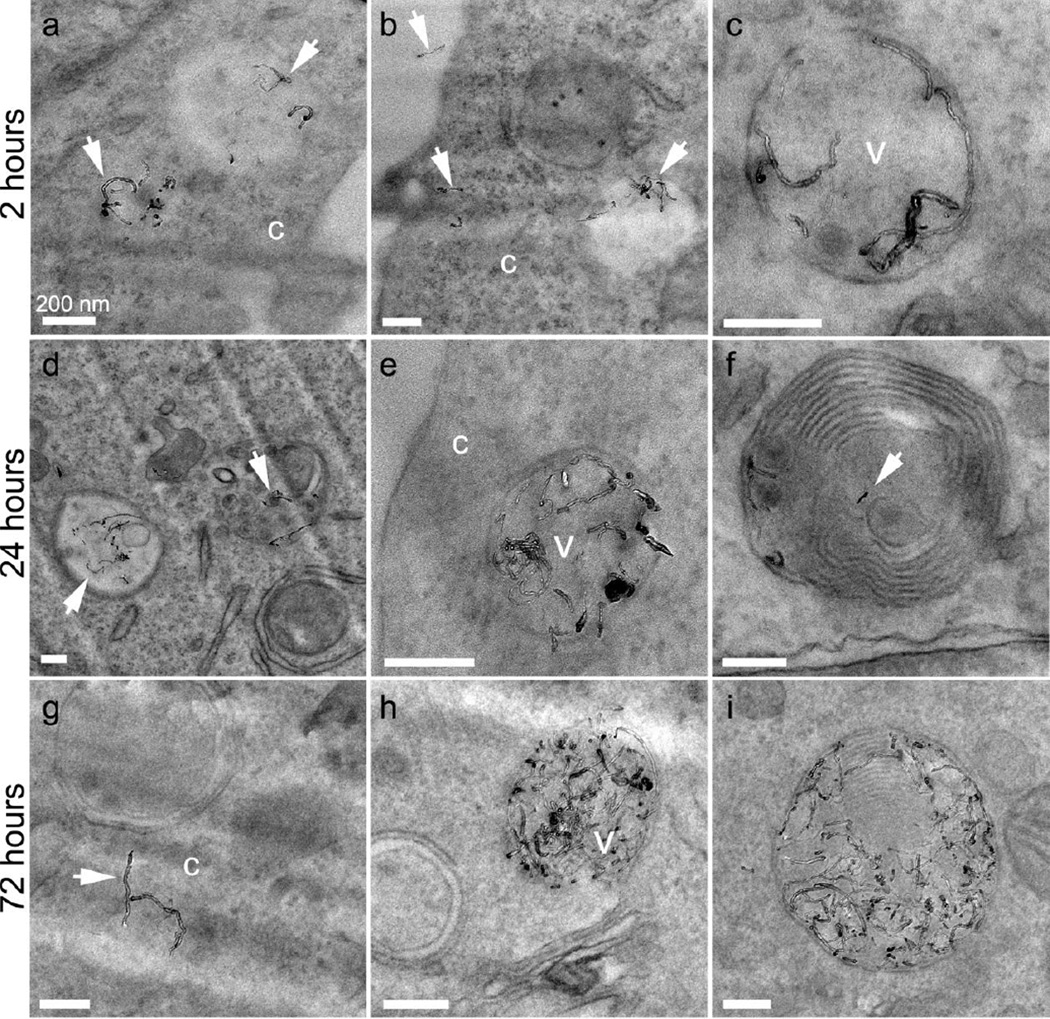
Figure 6.
TEM micrographs showing 20AO MWNTs internalized by N9 microglia after 2 (ac), 24 (d–f) and 72 (g–i) hrs. Selected MWNTs are marked with white arrows, and can be observed individually and in small clusters within vesicles (v) as well as in the cell cytoplasm (c). At longer time-points, multilaminar bodies are also observed containing MWNTs (f, i). All scale bars 200 nm.

