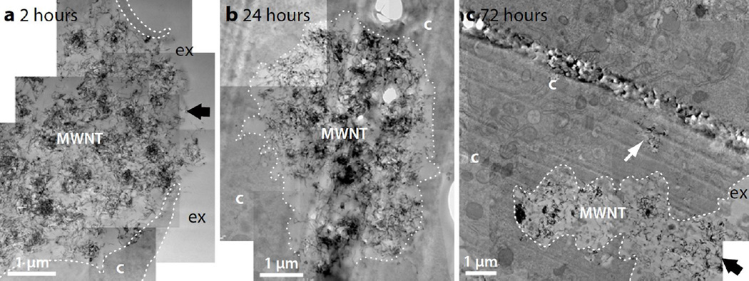
Figure 9.
TEM micrographs of N9 microglia exposed to pristine MWNT (10 µg/mL) after (a) 2 hour-, (b) 24 hour- and (c) 72 hour chase. Pristine MWNTs are present as large aggregates (labelled MWNT) and are often incompletely internalised by N9 microglia (black arrows), even after 72 hours. Cellular membranes are indicated with white dotted lines to more clearly distinguish the cytoplasm (c) from the extracellular space (ex). An internalised aggregate of pristine MWNTs is indicated by the white arrow.

