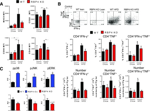
Figure 2.
Deletion of RBP4 reduces CD4 T-cell activation and AT macrophage JNK, Erk, and p38 phosphorylation. A: Expression of MHCII and costimulatory molecules (CD80, CD86, and CD40) in AT macrophages (CD11b+F4/80+). B: Flow cytometry representation (upper panel) and percentage (bottom panel) of intracellularly stained IFN-γ+ and TNF+ AT CD4 T cells. (n = 8 mice/group.) C: Phosphorylated (p)-p38, p-JNK, and p-Erk levels normalized to isotype control antibody in AT C45+CD11b+ cells from RBP4-Ox, RBP4 KO, and WT mice. (n = 4 mice/group.) Male mice, 23 weeks of age on HFD for 18 weeks for RBP4 KO and 18 weeks of age on chow diet for RBP4-Ox. Values are means ± SEM. *P < 0.05 vs. all other groups or as indicated. #P < 0.05 vs. chow-fed mice, same genotype. AU, arbitrary units; MFI, median fluorescence intensity; Pg, perigonadal.

