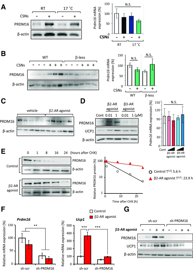
Figure 5.
Capsinoids (CSNs) stimulate a stabilization of the PRDM16 protein through the β2-AR pathway. A: Endogenous PRDM16 protein and relative mRNA expression of Prdm16 in the inguinal WAT of mice kept under ambient temperature (RT) or 17°C (n = 6). Mice were fed an HFD supplemented with 0.3% CSNs (+) or vehicle (CSNs [−]) for 8 weeks. β-Actin was used as a loading control for Western blotting. The Prdm16 mRNA expression data are mean ± SEM. B: Endogenous PRDM16 protein and relative mRNA expression of Prdm16 in the inguinal WAT of WT mice and β-less mice under 17°C (n = 6). Mice were fed an HFD supplemented with 0.3% CSNs (+) or vehicle (CSNs [−]) for 4 weeks. β-Actin was used as a loading control for Western blotting. C: PRDM16 protein expression was analyzed by Western blotting in the inguinal WAT of mice injected with vehicle (saline) or β2-AR agonist (formoterol) at a dose of 1 mg/kg/day for 1 week (n = 5). β-Actin was used as a loading control. D: Protein expression of PRDM16 and UCP1 was analyzed by Western blotting in primary inguinal WAT–derived adipocytes. The cells were cultured in the absence or presence of β2-AR agonist (formoterol) or β3-AR agonist (CL316243) at doses of 0.01 and 1 μmol/L throughout adipocyte differentiation (n = 3). β-Actin was used as a loading control. Relative Prdm16 mRNA expression in these cells is also shown. E: Cycloheximide (CHX) chase experiment in primary inguinal WAT–derived adipocytes. Endogenous PRDM16 protein expression was analyzed by Western blotting. Differentiated adipocytes were treated with vehicle or β2-AR agonist (formoterol) at a dose of 1 μmol/L in the presence of CHX for 1, 8, 16, and 24 h. β-Actin was used as a loading control. Regression analysis of PRDM16 protein stability is also shown. F: Relative mRNA expression of Prdm16 and Ucp1 was measured by qRT-PCR in primary inguinal WAT–derived adipocytes infected with sh-scr or sh-PRDM16 (n = 3). The adipocytes were treated with vehicle (control) and β2-AR agonist (formoterol) at a dose of 1 μmol/L throughout adipocyte differentiation. **P < 0.01, ***P < 0.001. G: PRDM16 and UCP1 protein expression was analyzed by Western blotting in the primary inguinal WAT–derived adipocytes shown in panel F. β-Actin was used as a loading control. Cont, control; N.S., not significant; T1/2, half-life.

