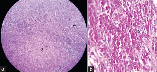
Figure 3.

(a) Histopathology combined feature of malakoplakia and xanthogranulomatous pyelonephritis in same slide. Arrow head: Granuomatous lesions. Star: Sheets of histiocytes (H and E, ×40). (b) Histopathology showing Michaelis–Gutmann body (arrow head) with foamy histiocytes (PAS, ×400)
