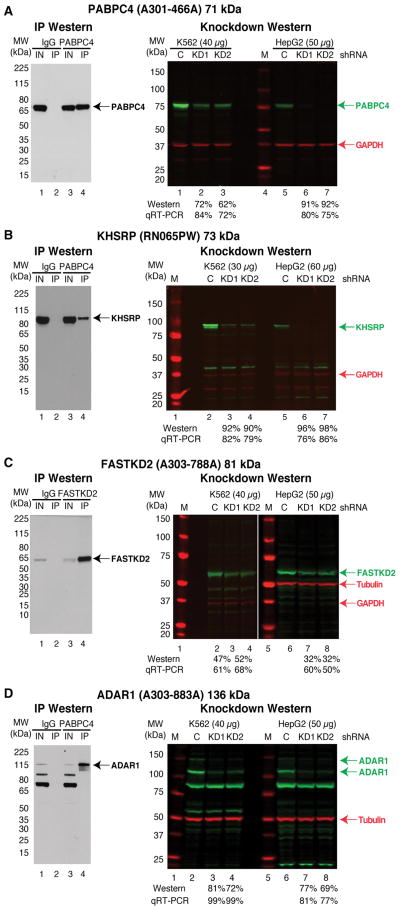
Figure 4. shRNA knockdown-western blot validation of antibodies against RBPs.
Representative IP-WB (left) and shRNA knockdown-western blot validations (right) for antibodies that cover a spectrum of band patterns. Experiments are shown for antibodies that recognize PABPC4 (A), KHSRP (B), FASTKD2 (C), and ADAR1 (D). For each experiment the molecular weight markers are shown along with the percent depletion in the knockdown sample compared to the control shRNA sample for both the western blot and qRT-PCR experiments. The position of the RBP (green) and the loading controls of GAPDH or Tubulin (red) are shown. See also Table S3 and Table S5.

