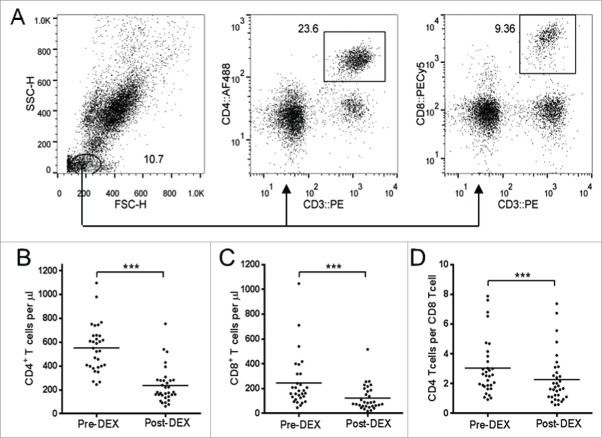
Figure 2.
Dex treatment reduces the number circulating of CD4+ and CD8+ T cells. (A) Representative flow cytometry data showing gating strategy on whole blood samples used to obtain absolute volumetric cell count data. Lymphocytes were identified on the basis of forward scatter (FSC) vs. side scatter (SSC), with CD4+ or CD8 T cells were subsequently identified as CD3+CD4+ and CD3+CD8+ respectively. (B–D) Analysis of T cell subsets in peripheral blood samples collected before (pre-Dex) and after (post-Dex) administration of dexamethasone. (B) Concentration of CD3+CD4+CD8− lymphocytes per μL of peripheral whole blood. (C) Concentration of CD3+CD4−CD8+ lymphocytes per μL of whole blood. (D) The CD4:CD8 ratio, calculated by dividing the number of CD4+ T cells per μL by the number of CD8+ T cells per μL. Each dot represents an individual patient; significant difference between pre-Dex and post-Dex values: ***p < 0.0001, paired students t-test.

