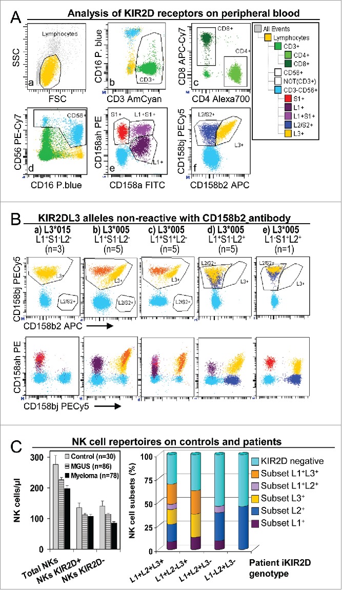
Figure 1.

NK cell repertoires in peripheral blood of controls and patients. (A) Analysis of KIR2D expression in peripheral blood NK and T cells. Gates to identify total lymphocyte (a), as well as CD3+ (b), CD4+ and CD8+ (c), CD16+CD56+ (d), KIR2DS1+, L1+ and L1+S1+ (e), and KIR2DL2/S2+ and L3+ (f) subsets were set in appropriate dotplots.35 These gates were logically combined to assigned KIR2D expression to CD3+CD4+, CD3+CD8+ and CD3−CD16+CD56+ (NK) lymphocytes in a representative patient with the KIR2DL1+S1+L2/S2+L3+ genotype. (B) Special analyses were implemented for patients with KIR2DL3 alleles that were non-reactive with anti-KIR2DL3 antibody clone 180701 (presumably KIR2DL3*005 or *015 alleles).36 As described, KIR2DL3*005 was cross-reactive with anti-KIR2DL1/S1 antibody clone EB6 but not with KIR2DL1 antibody clone 143211. In these patients, the possibility of three genetic backgrounds required cytometric analysis to be adapted: (1) for KIR2DL2− individuals (a, b and c; n = 13) all cells stained with CD158b/j (clone GL183) were assigned as L3+ (yellow and orange), disregarding alleged-alleles *005 and/or *015 were homozygous (a, n = 3 ), or heterozygous with others KIR2DL3 alleles (orange) which are reactive with the CD158b antibody (b, n = 5 and c, n = 5); (2) for KIR2DL2+ individuals (d and e; n = 6) cells co-reacting with anti-CD158b/j and CD158a/h antibodies were assigned as L3+ (yellow), and cells that were non-reactive with CD158a/h antibody as L2+ (dark blue); (3) for KIR2DS1+/KIR2DL3*005 individuals (c and e; n = 6 ), S1+ cells were considered only as those that reacted with CD158a/h but not with CD158a nor CD158bj antibodies (violet and red, in c and e, respectively). (C) NK cell repertoires in peripheral blood of controls and patients. Left, no differences in the number (cells/μL) of total NK cells (CD3−CD16+CD56+) or NK cells expressing KIR2D receptors (KIR2D+) or not (KIR2D−) between controls and MGUS or myeloma (SMM+MM) patients were detected by using CD158a/h+CD158bj antibodies. Right, distribution of peripheral blood NK cell subsets (percent of total NK cells) expressing KIR2DL1, and/or KIR2DL2/S2 and/or KIR2DL3, in total patients (MGUS+SMM+MM) with different KIR2D genotypes. KIR2DL1−L2+L3− showed only KIR2DL− or KIR2DL2+ clones.
