Fig. 4.
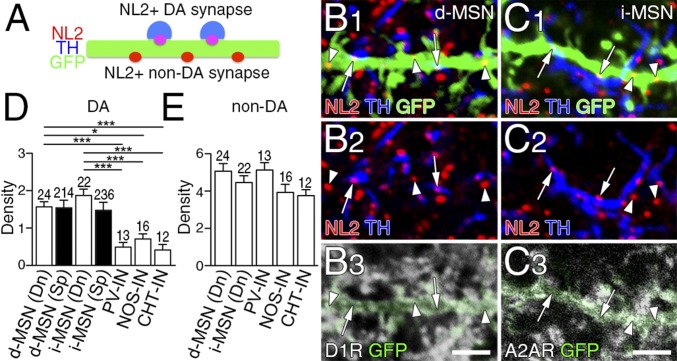
Dopamine synapses are preferentially formed on to dendrites of two types of MSNs. (A) Schematic to distinguish NL2-clustered dopamine and non-dopamine synapses on GFP-labeled dendrites of striatal neurons. (B and C) Quadruple immunofluorescence for NL2 (red), GFP (green), and TH (blue), and for D1R (B, white) or A2AR (C, white). NL2-clustered dopamine synapses (arrows) are preferentially distributed on dendrites of D1R-labeled d-MSNs (B) and A2AR-labeled i-MSNs (C). (D and E) The density of NL2-clustered dopamine (D) and non-dopamine (E) synapses per 10 μm of dendritic shafts (Dn) and spines (Sp) in d-MSNs and i-MSNs, and dendrites of striatal interneurons. Immunofluorescence images for striatal interneurons are shown in Fig. S4. The number of dendrites analyzed is indicated above each column. Error bars represent SEM. *P < 0.05 and ***P < 0.001 (one-way ANOVA with Tukey’s post hoc test). (Scale bars, 2 μm.)

