Fig. 5.
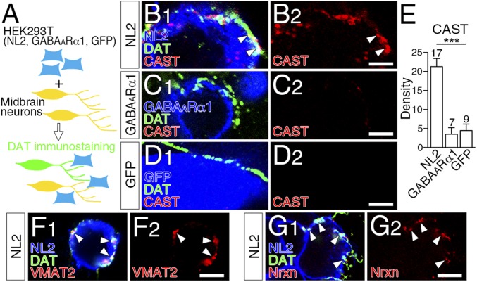
NL2-mediated presynaptic differentiation of dopaminergic axons in vitro. (A) Schematic of coculture assay of midbrain neurons with HEK293T cells expressing NL2, GABAARα1, or GFP. Axons of midbrain dopamine neurons were identified by DAT immunofluorescence. (B–D) Triple immunofluorescence for CAST (red) and DAT (green), and for NL2 (B, blue), GABAARα1 (C, blue) or GFP (D, blue). CAST clusters are recruited to contact sites of dopaminergic axons with HEK293T cells expressing NL2 (B, arrowheads), but not GABAARα1 (C) or GFP (D). (E) The density of CAST clusters per 100 μm of dopaminergic axon in contact with HEK293T cells. The number of HEK293T cells contacted by DAT-labeled dopaminergic axons is indicated above each column. Error bars represent SEM. ***P < 0.001 (Mann–Whitney u test). (F and G) Triple immunofluorescence for VMAT2 (F, red) or Nrxn (G, red), DAT (green), and NL2 (blue) in cocultures of midbrain dopamine neurons and HEK293T cells expressing NL2. (Scale bars, 2 μm.)

