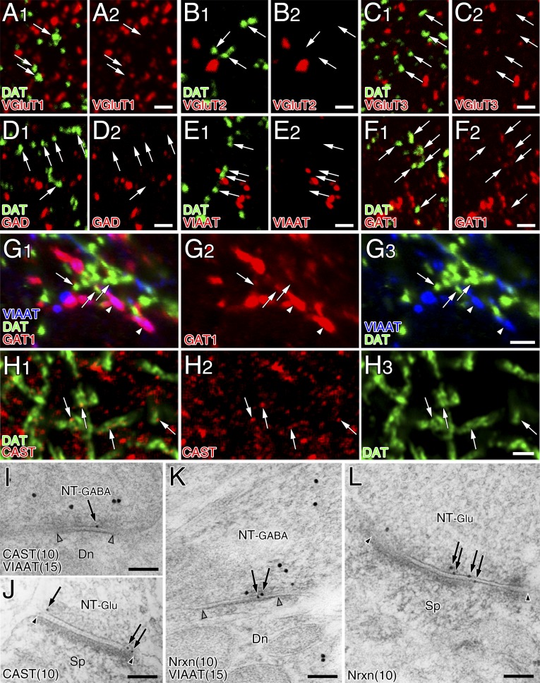
Fig. S1.
Presynaptic phenotype at striatal dopamine synapses is neither glutamatergic nor GABAergic. (A–F) Double immunofluorescence for DAT (green) and terminal markers (red), including VGluT1 (A), VGluT2 (B), VGluT3 (C), GAD (D), VIAAT (E), or GAT1 (F). Ultrathin (100 nm) sections were used to increase the spatial resolution of fluorescent signals. Note the lack of glutamatergic and GABAergic molecular expression in DAT+ dopaminergic terminals, except for weak immunoreactivity for GAT1 (arrows in F). (G) Triple immunofluorescence for DAT (green), GAT1 (red), and VIAAT (blue) showing much stronger expression of GAT1 in VIAAT-labeled GABAergic terminals (arrowheads) than in DAT-labeled dopaminergic terminals (arrows). (H) Superresolution immunofluorescence images for CAST (red) and DAT (green). Tiny CAST+ puncta are detected in some portions of large DAT+ dopaminergic terminals (arrows). (I–L) Double-label postembedding immunoelectron microscopy for VIAAT [Ø (diameter) = 15-nm colloidal gold particles] and CAST (I and J, Ø = 10 nm) or Nrxn (K and L, Ø = 10 nm). Immunogold labeling for CAST and Nrxn is observed beneath the presynaptic membrane at VIAAT+ symmetric or GABAergic synapses (NT-GABA, I and K) and VIAAT− asymmetric or glutamatergic synapses (NT-Glu, J and L). Pairs of open and filled arrowheads indicate the synaptic membrane of symmetric and asymmetric synapses, respectively. Dn, dendrite; Sp, spine. [Scale bars: 2 μm (A–H) and 100 nm (I–L).]

