Fig. 5.
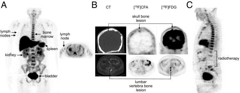
[18F]CFA PET CT images in humans. (A) Images of [18F]CFA biodistribution in a healthy volunteer. Maximum intensity 2D projection whole body image (Left) and cross-sectional tomographic image (Right) of the axilla indicating probe accumulation in the lymph nodes. (B) [18F]CFA PET/CT and [18F]FDG PET/CT images of a paraganglioma patient showing variability in [18F]CFA accumulation between the skull lesion and the vertebra lesion. (C) Whole body [18F]CFA image of the same patient as in B, showing reduced probe accumulation in previously irradiated lumbar spine region.

