
Fig. 4.
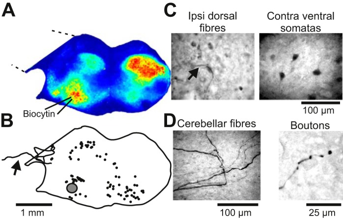
Retrograde labeling of neurons following local co-iontophoresis of biocytin and KCl in roller-drum cultures. A: oscillatory calcium activity (Z projection of standard deviation from 300-frame image stack) in a roller-drum brain stem-cerebellar coculture. B: camera lucida reconstruction of the position of retrogradely labeled neurons (solid circles) after local co-iontophoresis at the ventral oscillatory group (shaded circle). Note that some labeled fibers cross into the cerebellar explant (arrow). C: photomicrograph of labeled fibers located in the ipsilateral dorsal part, and somata in the contralateral ventral part, of the coculture. D: photomicrograph of labeled fibers entering the cerebellar part of the cultures and ending in boutons.