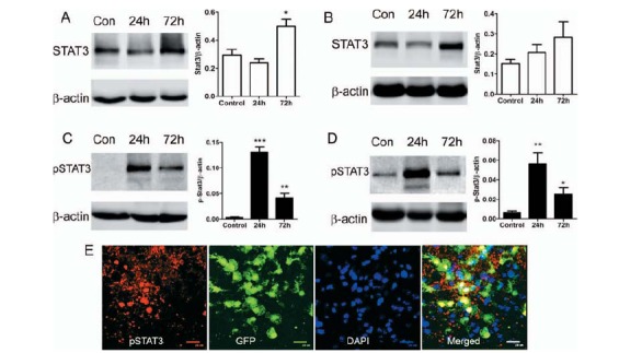
Fig. (7).

The expression of STAT3, pSTAT3 in the retina and RPE/choroid in laser-induced CNV. Laser-induced CNV was conducted in WT C57BL/6 mice. Eyes were collected at 24h and 72h after and retina (A, C) and RPE/choroid (B, D) were processed for Western Blotting analysis of STAT3 (A, B) and pSTAT3 (C, D). β-actin was used as housekeeping reference protein. Mean ± SD, n = 5. *, P < 0.05, **, P < 0.01, ***, P < 0.001 compared to non-CNV controls. One-Way ANOVA followed by Tukey’s Multiple Comparison Test. E, Laser-induced CNV was conducted in CX3CR1gfp/+ mice. 24h later, eyes were collected for confocal flatmount investigation of pSTAT3 expression. A confocal image of RPE/choroidal flatmount showing pSTAT3 expression in CNV lesion and infiltrating CX3CR1+ cells.
