Figure 3.
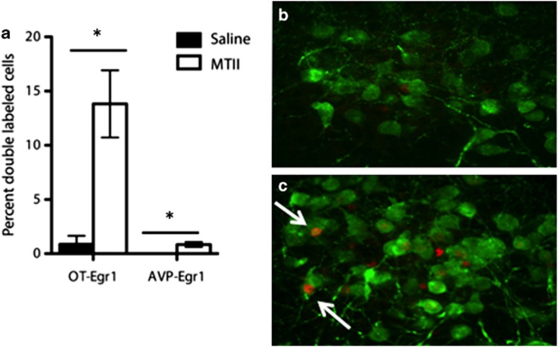
MTII activates OT-positive neurons in the paraventricular nucleus. (a) In the paraventricular nucleus of the hypothalamus, 13.8% of OT-positive cells were activated by in vivo MTII administration, as indicated by EGR1 expression, compared with <1% of cells under control conditions (p<0.05). Less than 1% of vasopressin-positive cell were activated by MTII administration. (b) OT-positive paraventricular cells after saline administration (green=OT, red=EGR1). (c) OT-positive cells after MTII administration. Arrows indicate colocalization of OT and EGR1. *Indicates a significant difference in the proportion of double-labeled neurons (p<0.05).

