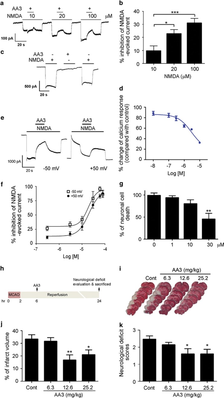
Figure 3.
Anemoside A3 (AA3) protects against NMDA receptor (NMDAR)-mediated excitotoxicity through the direct modulation of NMDAR. (a–f) AA3 is a non-competitive NMDAR modulator. (a) Current traces and (b) quantitative analysis of AA3-dependent inhibition of NMDA-evoked current in hippocampal neurons at a holding potential of −50 mV. Inhibition of NMDA-evoked current by AA3 increased with increasing concentrations of NMDA. n=4–5, *p<0.05, ***p<0.005 vs 10 μM. (c) The presence of NMDA (200 μM) was required for the blockade effect of AA3 (30 μM) on NMDAR. Current traces are shown. (d) AA3 inhibited the NMDA-induced calcium influx in cultured hippocampal neurons. (e) Current traces showing the blockade effect of AA3 on NMDA-evoked current at holding potentials of −50 and +50 mV. (f) Concentration–response curves showing the inhibition of NMDA-evoked current by AA3 at holding potentials of −50 and +50 mV. n=5–11. (g) AA3 protected cultured hippocampal neurons from NMDA-induced excitotoxicity (200 μM). n=4 experiments, **p<0.01 vs 0 μM. (h–k) AA3 protected against ischemic brain injury in adult rats. (h) Schematic diagram illustrates the time line of AA3 administration and tissue collection in middle cerebral artery occlusion (MCAO). AA3 was orally administered to rats at 6 h after MCAO. (i) Representative images of brain slices stained with TTC. TTC-stained red regions indicate unaffected tissue and pale white regions show infarcted tissue. AA3 reduced infarct volume (j) and neurological deficit scores (k) in rats under ischemic injury. n=8–10, *p<0.05, **p<0.01 vs control (Cont).

