Figure 1.
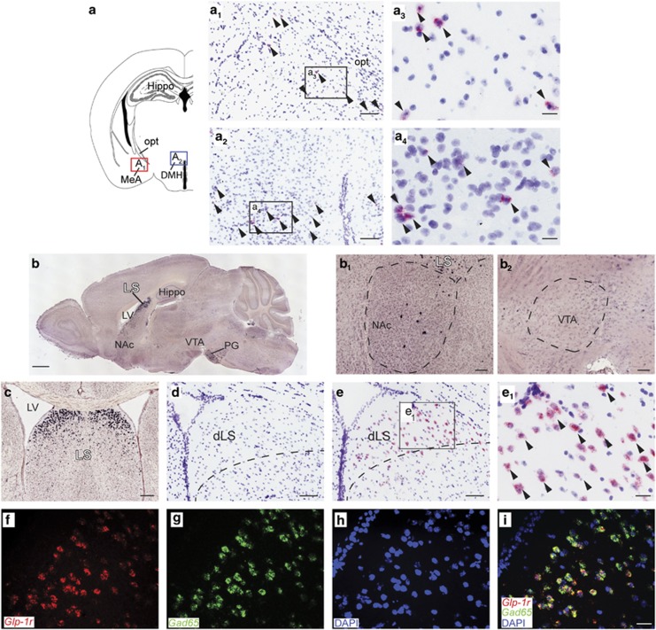
Glp-1r mRNA is enriched in the lateral septum. (a) Illustration of a coronal mouse brain section. (a1, a2) Glp-1r mRNA-expressing cells (purple) in the medial amygdaloid nucleus (red box in (a)) or the hypothalamus (blue box in (a)) labeled by arrowheads. (a3, a4) High magnification of individual Glp-1r mRNA-positive cells. (b) Overview of Glp-1r mRNA expression (black staining) domains in the sagittal plane (Allen Brain Atlas ID 73606497_125). (b1, b2) Close-ups of the NAc and the VTA showing little Glp-1r expression. (c) In the coronal plane, highest Glp-1r expression (silver impregnation) was identified in the LS (Allen Brain Atlas ID 74511737_345). (d) The Glp-1r sense probe produced no signal. (e) Abundant Glp-1r mRNA expression (pink) was confirmed in the dLS using a specific DIG-labeled riboprobe. (e1) Higher magnification of the dLS indicated by the box in (e) depicting strong cellular signal detection of Glp-1r mRNA. (f) Fluorescent detection of Glp-1r (red) and (g) Gad65 (green) mRNA in the dLS. (h) Nuclear counterstaining with DAPI (blue). (i) Merged picture reveals that almost all Glp-1r-positive septal neurons are GABAergic. Bars: (a1, a2, d, e) 75 μm; (a3, a4, e, f) 10 μm; (b) 1 mm; (b1, b2) 100 μm; (c) 200 μm. dLS, dorsal lateral septum; DMH, dorsomedial hypothalamic nucleus; Hippo, hippocampus; LV, lateral ventricle; MeA, medial amygdaloid nucleus; NAc, nucleus accumbens; opt, optic tract; PG, pontine gray; VTA, ventral tegmental area.

