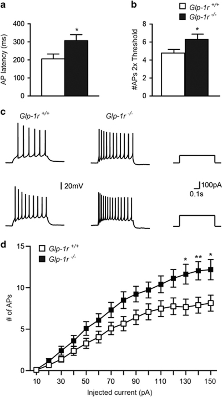
Figure 2.
Increased firing of Glp-1r−/− septal neurons. (a) The time to fire an AP is delayed in Glp-1r−/− (total of 23 cells from 10 animals) compared with Glp-1r+/+ (total of 21 cells from 11 animals) neurons (P=0.02). (b) The number of APs at twice threshold was elevated in Glp-1r−/− compared with Glp-1r+/+ controls (P=0.04). (c) Representative responses of Glp-1r+/+ (left) and Glp-1r−/− (right) neuron, respectively, to 600 ms depolarizing current steps at 100 and 150 pA. (d) Responses (number of APs evoked by 600 ms stimulus) of Glp-1r+/+ and Glp-1r−/− cells across a range of step current injections from 10 to 150 pA revealed an increased activity of Glp-1r−/− neurons (two-way repeated measures ANOVA × genotype effect: F(1, 43)=6.48, P=0.01). *P<0.05, **P<0.01.

