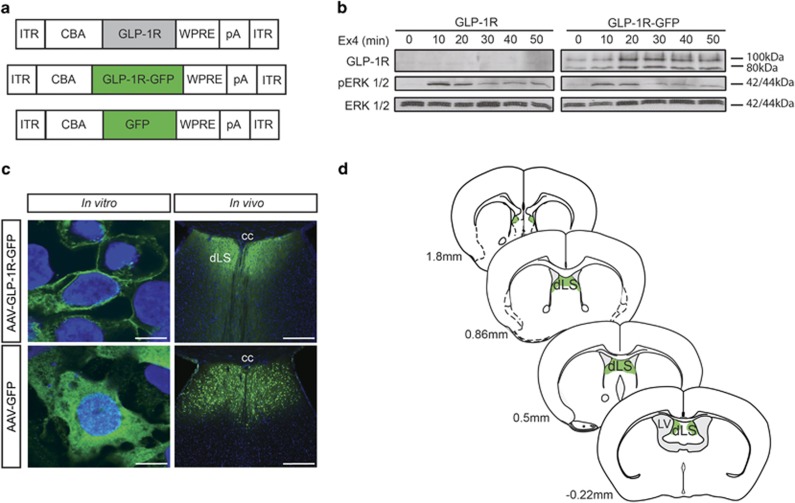
Figure 3.
AAV-mediated Glp-1r gene delivery to the dLS. (a) AAV expression cassettes. ITR, inverted terminal repeat; CBA, chicken β-actin promoter; pA, polyadenylation signal; WPRE, woodchuck hepatitis virus posttranscriptional regulatory element. (b) Biological activity of GLP-1R-GFP was confirmed by expression in heterologous HEK293 cells and exposure to Ex4 (10 nM) for the indicated durations (0–50 min). The dynamics of ERK phosphorylation were similar following activation of the tagged GLP-1R or the native receptor. Total ERK served as loading control. Note that the GFP antibody exclusively recognized the tagged recombinant receptor. (c) Recombinant GLP-1R-GFP traffics to the cell surface in HEK293 cells and in the mouse LS (top row), whereas GFP controls show cytosolic expression (bottom row). Anti-GFP immunofluorescence (green); DAPI (blue) nuclear counterstain. Bars: 10 μm. (d) Schematic illustration of vector spread assessed by AAV-transduced neuronal somata; cc, corpus callosum; dLS, dorsal lateral septum; LV, lateral ventricle.

