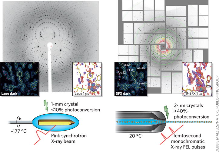
Figure 3.

Comparison of time-resolved Laue crystallography and time-resolved SFX crystallography. Left, Laue crystallography; right, time-resolved SFX. Examples are shown here for PYP. The diffraction patterns and the crystal photo were kindly provided by M. Schmidt. Electron density data are reproduced with permission from ref. 18.
