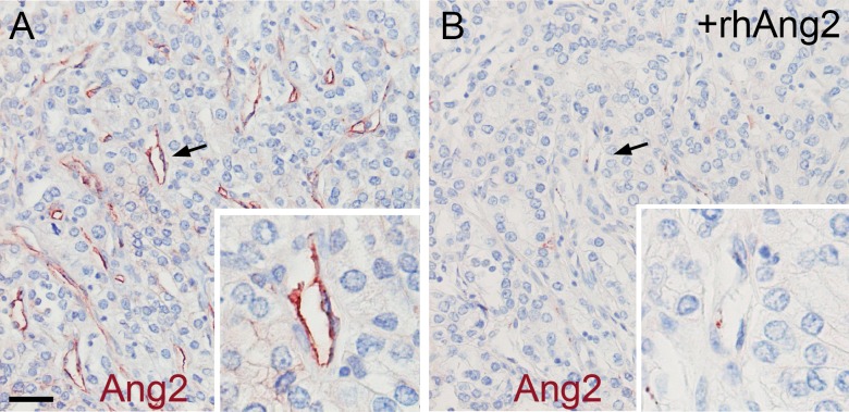
Fig 1. Representative immunohistochemical staining of Ang2 in mRCC.
(A) immunohistochemical staining of paraffin-embedded mRCC primary tumour tissue using a polyclonal antibody to Ang2. The CD31 expression was scored 3 in the sample. (B) An adjacent section was stained using the polyclonal antibody to Ang2, blocked with a 5-fold molar excess of recombinant human Ang2 (+rhAng2). Arrows indicate the magnified areas. Magnifications x200 and x400. Scale bar 40 μm.

