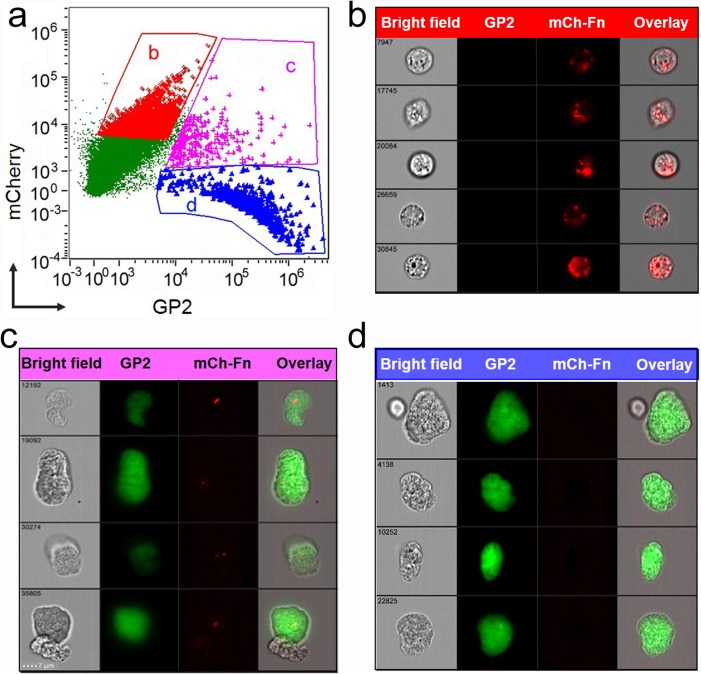
Fig 1. Uptake of ΔiglB vaccine strain by intestinal M-cells following oral inoculation.
BALB/c mice (4–6 wks) were orally vaccinated with mCherry-expressing ΔiglB (108 CFU/100 μL). Small intestines were collected at 90 min. post-vaccination and Peyer’s patches were excised to generate single cell suspensions. Cells were labeled with AF-488 conjugated α-GP2 antibody and subjected to cytometry imaging analysis. (a) Dot-plot depicts GP2 and mCherry intensity of each examined cell and three gated cell populations: (b) mCherryhiGP2low, (c) mCherryhiGP2hi, and (d) mCherrylowGP2hi. The representative cell images of these three gated populations are shown in (b) ΔiglB residing in non-M-cells, (c) ΔiglB residing in M-cells, and (d) M-cells with no ΔiglB, respectively. Representative images from 2 independent experiments are shown.

