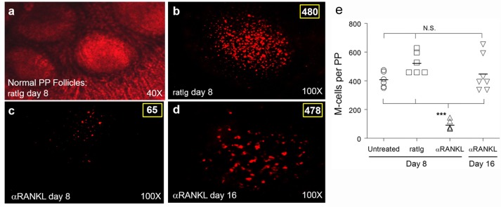
Fig 2. M-cells are depleted with αRANKL antibody treatment.
BALB/c mice (n = 3 per group) were treated i.p. with 250 μg of either rat Ig (a-b) or αRANKL antibody IK22-5 (c-d) on days 0, 2, 4, and 6. On day 8 (a-c) or day 16 (d), animals were sacrificed and Peyer's patches (PP) collected and stained with rhodamine-labeled UEA-1 for whole mount imaging. Representative images showing normal morphology of intact (ratIg-treated) PP were taken using combined phase contrast and fluorescence at 40x (a), or labeled M cells (480 in rat Ig-treated PP on day 8, 65 M-cells in αRANKL-treated PP on day 8, or 476 in αRANKL-treated PP on day 16) all with fluorescence alone at 100x (b-d). M-cells in PP whole mounts were quantified for naive, rat Ig-treated, and αRANKL-treated groups (e), with αRANKL treatment inducing a significant decrease in M-cells (***p < 0.001) compared to naive or rat Ig-treated animals at day 8 or αRANKL-treated animals upon PP repopulation (day 16). Representative images from 3 experiments are shown.

