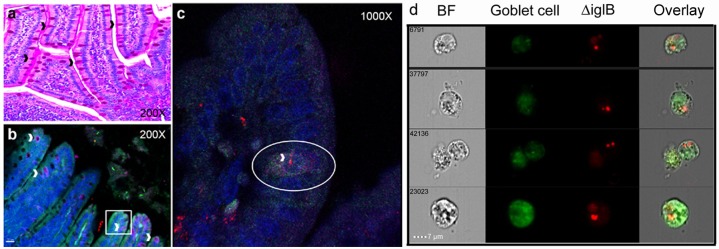
Fig 4. Goblet cells can take up Francisella tularensis by 90 minutes after oral vaccination.
BALB/c mice (n = 3) were orally vaccinated with mCherry-ΔiglB (approximately 108 CFU) and rested for 90 minutes prior to sacrifice for collection of whole intestines, which were paraffin embedded, sectioned, and stained for (a) periodic acid Schiff to visualize GCs (black arrowheads) and (b-c) confocal analysis with nuclear stain DAPI (blue), mucin marker anti-MUC-2 (green), and GC surface marker anti-cytokeratin-18 (pink), with GCs shown by white arrowheads. (c) High magnification of a goblet cell (circled), stained with both MUC-2 and cytokeratin-18, which has taken up ΔiglB (red). (d) Single cells were prepared from intestines of similarly vaccinated mice, labeled with FITC-anti-MUC-2 (green), and subjected to cytometry imaging to visualize the uptake of mCherry-ΔiglB (red) by goblet cells. BF, bright field. Representative images from 2 separate experiments are shown.

