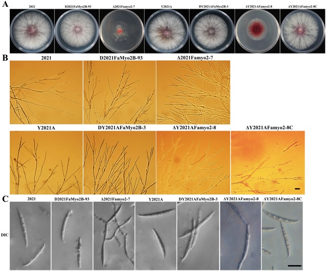
Fig 1. Colony and conidia morphology and hyphal tip growth and branching patterns of the wild-type 2021, the FaMyo2B disruption mutant, the Famyo2 deletion mutant and the Famyo2 complement mutant.
(A) Colonies were photographed after 3 days at 25°C on PDA. (B) The branching angles of the hyphae were reduced and distorted in the extension zone of the mutant colonies that had grown on a thin layer of water agar for 36 h. Bar = 200 μm. (C) Photographs of conidia obtained in the conidiation assay (see text). Bar = 20 μm.

