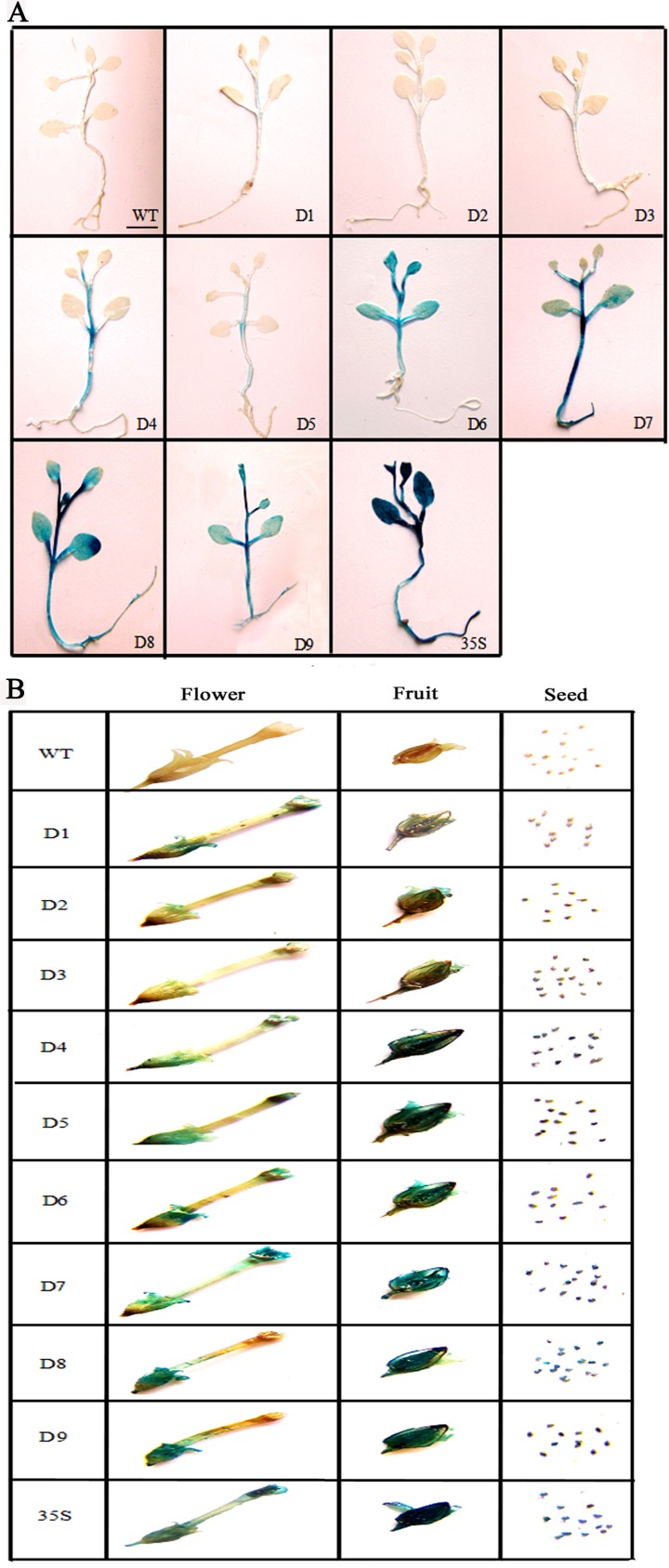
Fig 2. GUS histochemical assays of tissues of D1–D9 and CaMV 35S transgenic tobacco plants.
Twenty-day-old seedlings (A), and flowers, fruits and seeds (B) were incubated in staining solution at 37°C. The D1–D3 and D4–D9 fragments were stained for 24 h and 6 h, respectively, following which the samples were observed and photographed after decolorization. Scale bar: 0.5 cm.

