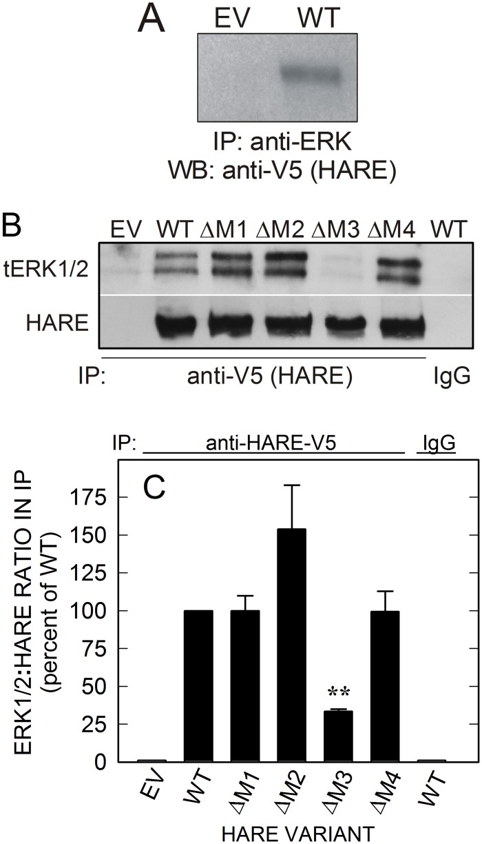
Fig 4. The HARE M3 motif is required to form complexes with ERK.
Cells stably expressing EV, WT or the indicated single-motif deletion HARE CD mutant were grown and processed as described in Methods and then incubated with or without 10 μg/ml (709 nM) UFH for 20 min and processed as in Fig 1. A. EV and WT cells were treated as in Fig 3 and Methods but in the absence of Hep or other ligands and cell lysates (300 μg of protein) were incubated with anti-tERK1/2 Ab. Immunoprecipitates were subjected to SDS-PAGE and Western blot analysis and blotted proteins were analyzed for HARE using anti-V5 Ab. B. HARE from equal amounts of lysate (300 μg of protein) from the indicated cells was incubated with goat anti-V5 Ab (2 μg/ml) and WT extract was also incubated with nonimmune control goat Ab (IgG). The immunoprecipitates were subjected to SDS-PAGE and Western blot analysis as described in Methods. Blotted proteins were analyzed using anti-tERK1/2 or anti-V5 Ab as indicated. C. Blots from independent experiments (n = 3), as in panel B, were scanned, digitized and densitometry analyses were performed to determine ERK:HARE ratios. The data are presented as mean ± SEM percent of the tERK:HARE ratio compared to WT as 100%. Samples with significant differences are indicated: **, p < 0.005.

