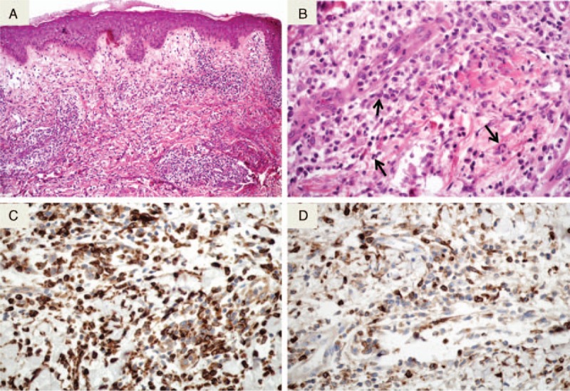
FIGURE 1.

Histological aspect of H-SS. A: As in the classical form of SS (N-SS), H-SS features edema of the papillary dermis and diffuse and interstitial dermal infiltrates (HES original magnification ×100). B: The infiltrate consists of lymphocytes and histiocytoid cells. Some cells are suggestive of myeloid progenitors (HES original magnification ×400). C: The infiltrate strongly express the CD68 monocyte marker (immunohistochemistry revealed by diaminobenzidine). D: A proportion of mononuclear cells express the myeloperoxidase (immunohistochemistry revealed by diaminobenzidine). SS = Sweet syndrome, HES = hematoxylin, eosin ans saffron, H-SS = histiocytoid Sweet syndrome, N-SS = neutrophilic Sweet syndrome.
