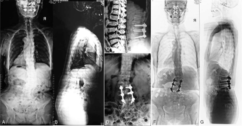
FIGURE 2.

(A/B) A 65-year-old female patient belonged to Group 2, with a lumbar curve as 30°, lumbar lordosis as 15°, thoracic lordosis as 5°. (C) Magnetic resonance imaging showed disc hernia occurred at L2/3 and L3/4. (D/E) The patient underwent decompression and 2 levels fusion (TLIF). (F/G) At 3-year follow-up, the balance was well maintained. TLIF = transforaminal lumbar intervertebral fusion.
