
FIGURE 2.
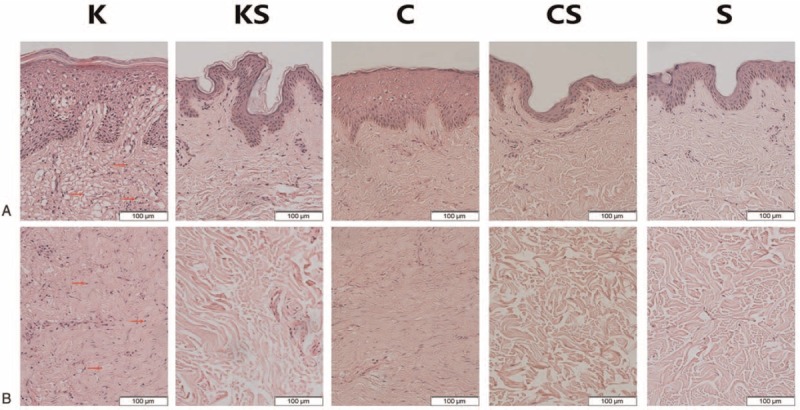
The results of H&E staining of epidermis (A) and dermis (B) in all groups. The number of infiltrated cells (red arrow) is much lower in the KS group, C group, CS group, and S group than in the K group (images: 200×). The epidermis of keloid and scar tissue is thicker than that of normal skin tissue. In keloid tissues, a large amount of fibroblasts with more cytoplasm and clear nucleoli are observed in the papillary layer and reticular layer of the dermis. Irregular, thick and extremely compact collagen fibrils appear disordered. By contrast, the nucleolus of normal skin tissue fibroblasts is small and there is less cytoplasm. In scar tissues, the collagen fibrils are relatively compact. Fibroblasts manifest more cytoplasm and a small nucleolus. C group = scar samples from normal scar patients, CS group = normal skin samples from scar patients, H&E = hematoxylin and eosin, K group = keloid samples from keloid patients, KS group = normal skin samples from keloid patients, S group = normal skin samples from patients without obvious scarring.