
FIGURE 1.
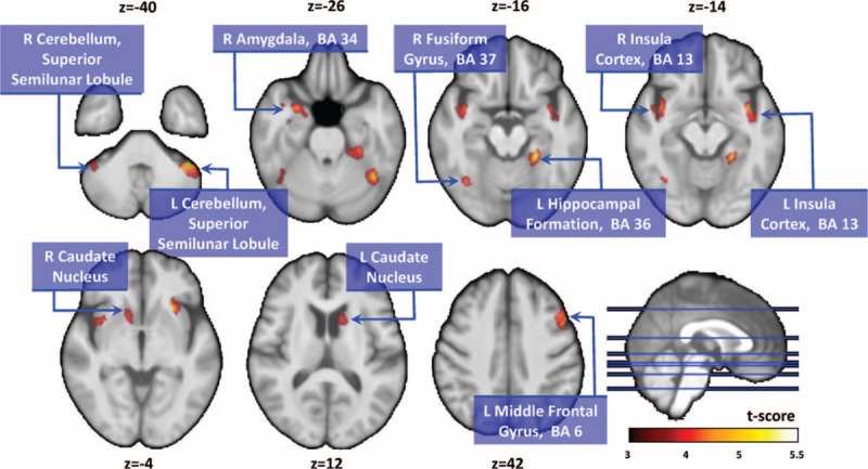
Compared with normal controls, patients with PD showed significantly smaller gray matter volumes in the right amygdala, left hippocampal formation, bilateral insular cortex, bilateral caudate nucleus, bilateral cerebellar superior semilunar lobule, right fusiform gyrus, and left middle frontal gyrus. PD = Parkinson disease.