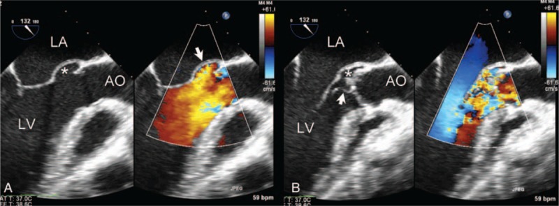
FIGURE 4.

Two-dimensional color Doppler transesophageal echocardiography showing P-MAIVF in the left ventricular outflow tract long-axis view. (A) Two-dimensional TEE showing P-MAIVF expanded (white star) and color Doppler imaging showing a flow filling the cavity with intact wall in systole. (B) P-MAIVF collapsed (white star), aortic vegetation (arrow), and severe aortic regurgitation in diastole. AO = aorta; LA = left atrium; LV = left ventricle; P-MAIVF = pseudoaneurysm of the mitral-aortic intervalvular fibrosa; TEE = three-dimensional transesophageal echocardiography.
