
FIGURE 3.
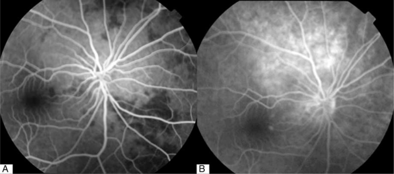
A, Fluorescein angiography of right eye (26.7 seconds after fluorescein injection). Note normal filling of retinal artery but several, fillings in the choroid. B, Fluorescein angiography of right eye (11 minutes and 42 seconds after fluorescein injection). Note late fluorescence leakage from choroid.