
Figure 6.
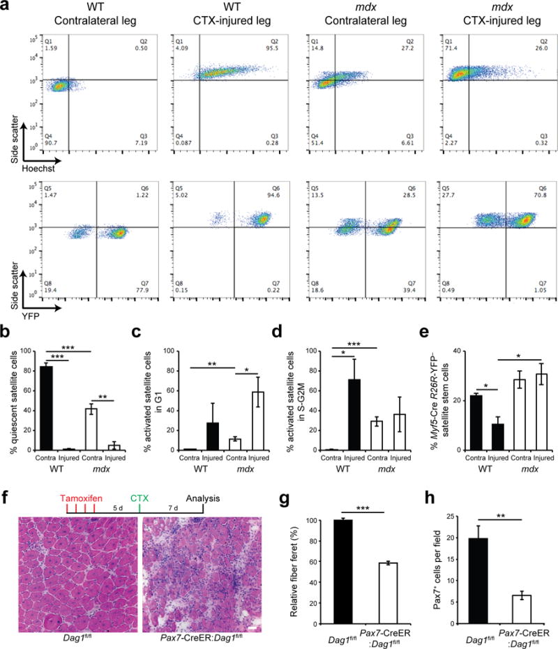
Dystrophin-deficient satellite cells have reduced ability to generate myogenic progenitors in regenerating muscle. (a) Flow cytometric analysis of WT and mdx Myf5-Cre:R26R-YFP mice 3 days after cardiotoxin (CTX) injury. Upper panels display the distribution of myogenic cells by side scatter (SSC, y-axis) and DNA content (x-axis) based on Hoechst 33342 staining, while lower panels profile their side scatter (y-axis) and YFP expression (x-axis). (b–d) Proportions of quiescent (SSC-low, DNA-low), activated G1 (SSC-high, DNA-low), and proliferating S-G2M (SSC-high, DNA-high) myogenic cells from CTX-injured or contralateral (Contra.) muscles of WT and mdx Myf5-Cre:R26R-YFP mice. (e) Proportions of YFP-negative satellite stem cells from CTX-injured or Contra. muscles of WT and mdx Myf5-Cre:R26R-YFP mice. (b–e) n = 3 mice per condition. (f) Representative pictures (n > 10 pictures per condition) from H&E staining of TA muscle sections of tamoxifen-treated Dag1fl/fl and Pax7-CreER:Dag1fl/fl mice 7 days after CTX-injury. n = 3 mice. Scale bar, 20 μm. (g) Relative fiber feret, and (h) satellite cell density (Pax7-expressing cells) from TA muscle sections of tamoxifen-treated Dag1fl/fl and Pax7-CreER:Dag1fl/fl mice 7 days after CTX-injury. n=3 mice per condition. (b–e,g,h) Error bars represent means ± SEM. *P < 0.05, **P < 0.01, ***P < 0.005. Statistical significance was calculated by Student’s t test.