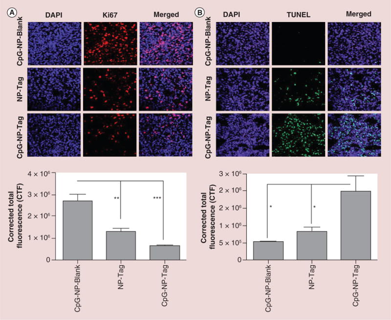
Figure 6. Effect of nanoparticle immunization on tumor cell proliferation and survival.

(A) Representative images (40×) of Ki67 stained tumor tissues sections showing rate of tumor proliferation and Quantitative analysis indicating proliferation rate of the tumor tissue harvested from the different groups analyzed using NIH ImageJ software (**p < 0.01; ***p < 0.001). (B) Representative images of TUNEL staining of the tumor sections showing the induction of apoptosis and Quantitative analysis of the apoptotic activity using TUNEL assay analyzed using NIH ImageJ software (*p < 0.05).
CTF: Corrected total fluorescence; DAPI: (4′,6-diamidino-2-phenylindole) fluorescent nuclear DNA stain; NP: Nanoparticle; Tag: Tumor antigen; TUNEL: terminal deoxynucleotidyltransferase (TdT)-mediated dUTP nick end labeling (TUNEL) assay.
