Fig. 3.
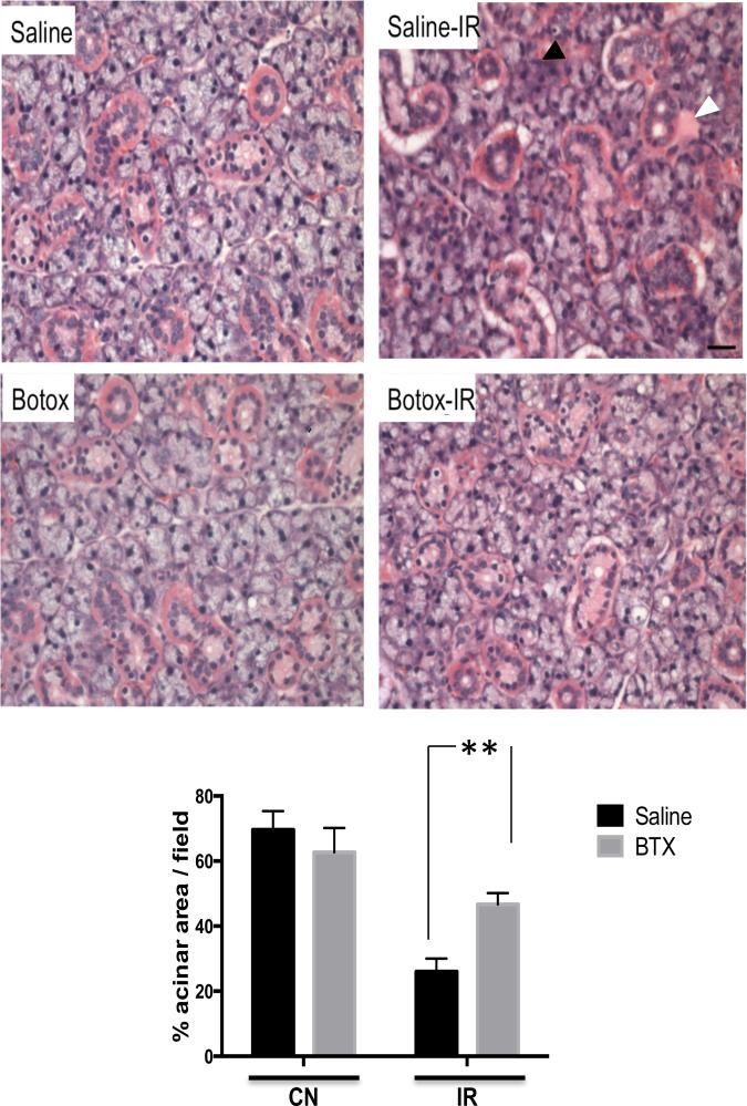
Botulinum toxin (Botox) injection mitigates irradiation (IR)-induced histologic changes. Tissue sections from submandibular glands were stained with hematoxylin and eosin at baseline and 4 weeks after radiation exposure. Degenerative changes with acinar atrophy (black arrowhead), periductal fibrosis (white arrowhead), and lymphocytic infiltrate are seen in the control (saline-IR) group. Botox-pretreated glands are markedly protected from acinar atrophy and periductal reaction (magnification ×40). Acinar area quantification revealed that botox preinjected glands contained significantly larger acinar areas compared with control (CN-saline) conditions; n=4/group, **P<.05.

