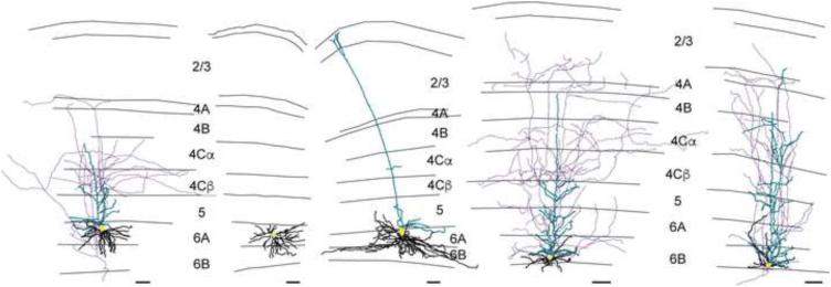
Figure 3.
Example V1 CG neurons of each type. Reconstructions of V1 CG neurons including (left to right) Iβ, spiny stellate, large, IC, and tilted cells. Apical dendrites are illustrated in turquoise, basal dendrites are illustrated in black, axons are illustrated in purple, and cell bodies are illustrated in yellow. Grey lines illustrate laminar borders and layers are labeled for all reconstructions except where layers on neighboring reconstructions are aligned. Scale bars beneath each reconstruction represent 100 microns. For additional reconstructions of each cell type, see Supplemental Figure 3.

