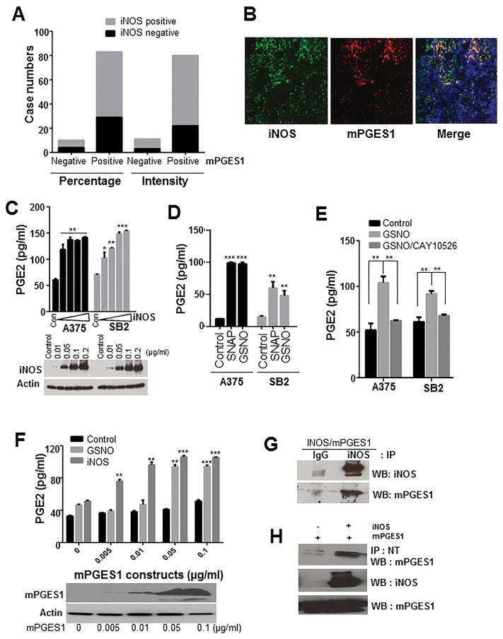
Figure 3. iNOS expression and NO donors regulate mPGES1 activity and PGE2 production in melanoma.
(A) Columns demonstrate N numbers of negative or positive staining of mPGES1 and iNOS. (B) A representative images of the staining of mPGES1 (red), iNOS (green) and DAPI (blue) in human melanoma specimens. (C) A375 and SB2 cells were transiently transfected with vector or iNOS construct. These cells were incubated in fresh medium for 2 days, and then iNOS expression, total nitrite, and PGE2 levels were determined. P <0.05 (*), P <0.001 (**), and P >0.001 (***) was considered statistically significant. (D) Cells were exposed to SNAP or GSNO (100 μM) for 24 h, and we performed an enzyme immunoassay to measure PGE2 levels. (E) Cells were pretreated with CAY10526 (5 μM) for 1 h, and then PGE2 levels were determined in cells exposed to GSNO (100 μM). (F) HEK293 cells were transfected with the indicated concentrations of mPGES1 construct and exposed to GSNO for 24 h or co-transfected with iNOS construct, and the PGE2 levels were measured. Actin and mPGES1 levels were determined using Western blotting. (G) HEK293T cells were co-transfected with iNOS and mPGES1 constructs. Then cell extracts were subjected to immunoprecipitation (IP) with immunoglobulin G (IgG) or anti-iNOS antibody, and the precipitates were analyzed by Western blot using the indicated antibodies (H) The extracts of HEK293T cells transfected with mPGES1 or mPGES1/iNOS constructs were subjected to IP with anti-nitrotyrosine antibody (NT), and mPGES1 levels were determined in the precipitates. Abbreviations: WB, Western blotting; IP, immunoprecipitation.

