
Figure 6.
Cervical cancer, stage IB2. Axial T2-weighted image shows an enlarged left internal obturator node with central necrosis (arrows). Caution should be taken to differentiate from normal ovary.
Official websites use .gov
A
.gov website belongs to an official
government organization in the United States.
Secure .gov websites use HTTPS
A lock (
) or https:// means you've safely
connected to the .gov website. Share sensitive
information only on official, secure websites.
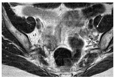
Cervical cancer, stage IB2. Axial T2-weighted image shows an enlarged left internal obturator node with central necrosis (arrows). Caution should be taken to differentiate from normal ovary.