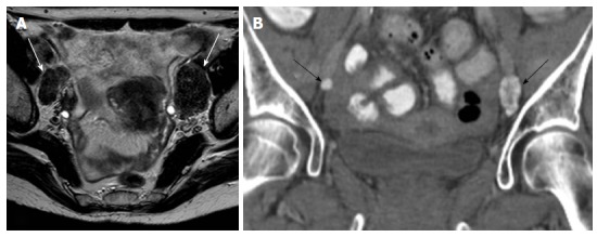
Figure 7.

Cervical cancer, stage IIB. A: Axial T2-weighted image shows bilateral enlarged external iliac lymph nodes with very low signal intensity (white arrows); B: Coronal reconstruction of computed tomography of the pelvis, confirms the presence of calcifications within the nodes (arrows). Calcification within metastatic nodes from cervical cancer is an unusual finding.
